Pain, stiffness, numbness or swelling in the hand and wrist can make everyday tasks such as writing, driving, opening jars or sleeping through the night surprisingly difficult. Most hand and wrist problems can be diagnosed clinically at a single consultation, and the majority are managed successfully without an operation.

My particular interest is the non-surgical management of hand and wrist pain, and in particular ultrasound-guided injection therapy for osteoarthritis and painful soft tissue conditions of the hand and wrist. I also assess and operate on ganglia and de Quervain’s tenosynovitis. Where an operation is needed for other conditions, such as carpal tunnel decompression, cubital tunnel surgery, trigger finger release or reconstruction for advanced arthritis, I work closely with specialist hand surgery colleagues and will arrange onward referral so that your care continues seamlessly.
 1
2
3
1
2
3
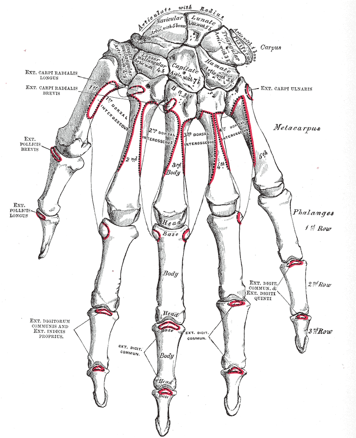
- 1Base of the thumb – the commonest site of hand arthritis
- 2The wrist and carpal tunnel – nerve compression and post-injury arthritis
- 3The finger joints – arthritis, and the pulleys that cause triggering
Use the quick reference list to jump straight to a condition, or tap any box to open the full information.
Your consultation
What happens at the appointment, and which tests are actually worth doing.
Click to find out moreClose
Your consultation
What happens at the appointment, and which tests are actually worth doing.
Assessment begins with a careful history of your symptoms, the activities that aggravate them and the impact they have on your work and daily life. Examination includes movement, strength, joint stability and specific nerve and tendon tests.
Investigations are requested only when they will change the treatment plan, and may include X-rays for arthritis, nerve conduction studies for suspected nerve compression, or diagnostic ultrasound, which can often be performed in clinic at the same visit.
Ultrasound-guided injections
Why guidance matters, what is injected, and what to expect in the days afterwards.
 Click to find out moreClose
Ultrasound-guided injections
Why guidance matters, what is injected, and what to expect in the days afterwards.
Injections are most effective when the medication is placed precisely where the problem lies. Ultrasound allows the joint, tendon sheath or cyst to be seen directly, the needle to be watched as it advances, and the injection to be confirmed as it is delivered. Compared with injecting by feel alone this improves accuracy, particularly in the small joints of the hand and around tendons, and it avoids injecting into the tendon itself.
Ultrasound also serves as a diagnostic tool at the same appointment. It shows thickening of the flexor and extensor tendon sheaths, swelling of the median nerve at the wrist, the size and origin of a ganglion, and the presence of joint inflammation or fluid. This means the diagnosis is confirmed and the treatment delivered in a single visit.
The injection itself usually contains a local anaesthetic together with a corticosteroid. For osteoarthritis, other options such as hyaluronic acid may be appropriate, and these are discussed on the injections pages of this site. Most injections take only a few minutes, and you can drive home afterwards unless you are told otherwise.
After an injection: what to expect
- The area may ache for one to three days as the local anaesthetic wears off before the steroid takes effect.
- Ice and simple painkillers are usually all that is required for this settling-in period.
- Benefit typically begins within a few days and continues to build over two to three weeks.
- Relief commonly lasts several months, and injections can be repeated at appropriate intervals.
- Temporary flushing, a small dimple or lightening of the skin at the injection site, and a short-lived rise in blood sugar in people with diabetes are recognised effects.
- Infection is rare but important; please make contact if the area becomes increasingly hot, swollen and painful after a few days, or if you feel feverish.
Osteoarthritis of the hand and wrist
Gradual loss of joint cartilage causing pain, stiffness, bony swelling and loss of grip.
 Click to find out moreClose
Osteoarthritis of the hand and wrist
Gradual loss of joint cartilage causing pain, stiffness, bony swelling and loss of grip.
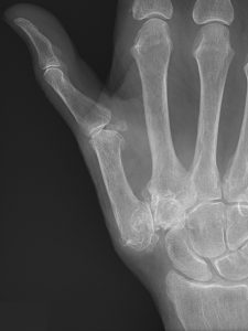
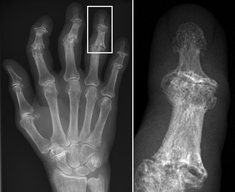
Osteoarthritis is the gradual loss of the smooth cartilage lining the joints, leading to pain, swelling, stiffness and deformity. In the hand it most commonly affects the joint at the base of the thumb, the small joints at the fingertips, the middle joints of the fingers, and the wrist, particularly after a previous fracture or ligament injury.
Typical symptoms
- Pain brought on by gripping, pinching, turning keys or opening jars
- Stiffness that is worst first thing in the morning and eases with gentle movement
- Bony swelling and enlargement of the finger joints
- Grinding or crunching sensations, and gradual weakness of grip and pinch
- Change in the shape of the thumb base, which may look squared off
How it is managed
The great majority of hand arthritis is managed successfully without surgery. Treatment is built around understanding the condition, pacing and adapting activities, and joint protection techniques, alongside simple aids such as thicker pens, lever taps, jar openers and padded handles. A custom thumb base or wrist splint reduces pain during aggravating tasks and can be worn at night. A hand therapy programme maintains movement and strengthens the muscles that stabilise the joints. Topical anti-inflammatory gels and simple analgesia are used according to your medical history.
Where these measures are not enough, an ultrasound-guided injection into the affected joint often gives several months of good relief and can be repeated at intervals. This is the mainstay of what I offer for hand and wrist arthritis, and for many patients it postpones or removes the need for surgery altogether.
If surgery is needed
If pain is no longer controlled and function is significantly limited, reconstructive surgery can be very effective. Options include trapeziectomy or joint replacement for the thumb base, and fusion or replacement of the finger joints, with a range of procedures available for the arthritic wrist. These operations are carried out by my hand surgery colleagues, and I will discuss whether referral is appropriate and arrange it for you.

Carpal tunnel syndrome
The median nerve compressed at the wrist, causing numbness, tingling and night-time symptoms.
 Click to find out moreClose
Carpal tunnel syndrome
The median nerve compressed at the wrist, causing numbness, tingling and night-time symptoms.
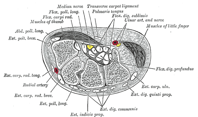
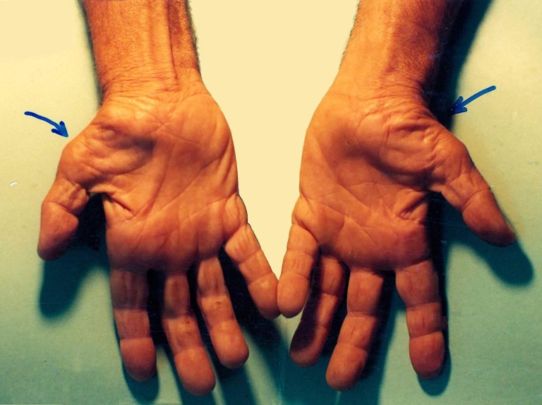
Carpal tunnel syndrome occurs when the median nerve is compressed as it passes through a tight fibrous tunnel at the front of the wrist. It is the most common nerve compression in the body and becomes more frequent with age, during pregnancy, and in people with diabetes, thyroid disease or inflammatory arthritis.
Typical symptoms
- Numbness, tingling or burning in the thumb, index, middle and half of the ring finger
- Symptoms that wake you at night and are relieved by hanging the hand out of bed or shaking it
- Clumsiness, dropping objects, or difficulty with fine tasks such as fastening buttons
- In longstanding cases, wasting of the muscle at the base of the thumb and permanent loss of sensation
Non-surgical management
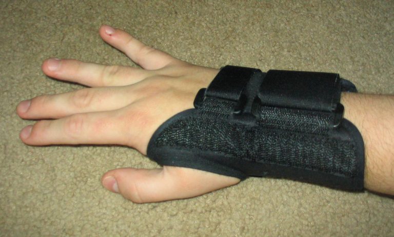
Mild or intermittent symptoms often improve with a night resting splint worn for six to eight weeks, activity modification, treatment of any underlying medical cause, and nerve gliding exercises with a hand therapist. An ultrasound-guided corticosteroid injection into the carpal tunnel can settle symptoms quickly, is a useful confirmation of the diagnosis, and may give relief lasting several months. Ultrasound guidance is particularly valuable here because it keeps the needle away from the nerve itself.
If surgery is needed
Carpal tunnel decompression is recommended when symptoms are persistent despite these measures, when they interfere with sleep or work, or when examination and nerve studies show progressive nerve damage. The operation divides the tight ligament forming the roof of the tunnel and is normally performed as a day case under local anaesthetic through a small incision in the palm. Night-time symptoms usually settle within days, and most people return to desk work within a few days and to heavy manual work after four to six weeks. This surgery is performed by my hand surgery colleagues, to whom I will refer you.

Cubital tunnel syndrome
The ulnar nerve compressed at the inner elbow, with numbness in the little and ring fingers.
 Click to find out moreClose
Cubital tunnel syndrome
The ulnar nerve compressed at the inner elbow, with numbness in the little and ring fingers.
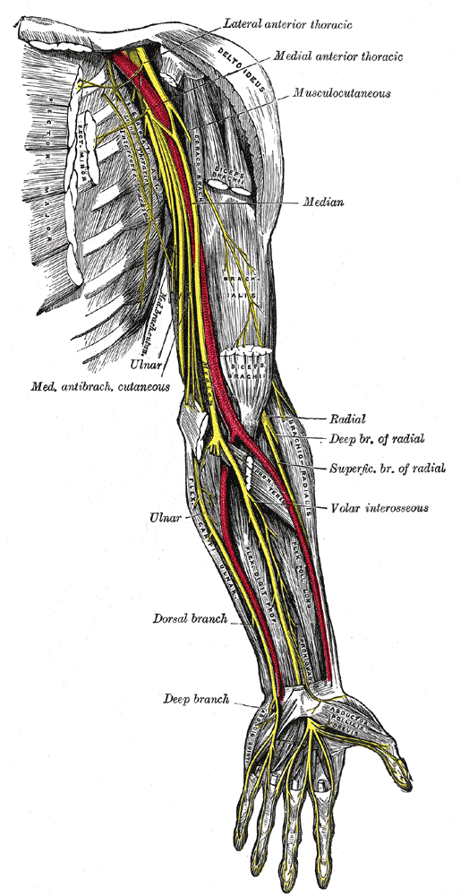
Cubital tunnel syndrome is compression of the ulnar nerve as it passes behind the inner side of the elbow. It is the second most common nerve compression in the upper limb and is often aggravated by prolonged elbow bending, leaning on the elbow, or previous elbow injury and arthritis.
Typical symptoms
- Pins and needles or numbness in the little finger and the adjacent half of the ring finger
- Aching along the inner elbow and forearm, sometimes radiating into the hand
- Weakness of grip and pinch, and difficulty crossing or splaying the fingers
- In advanced cases, visible wasting between the thumb and index finger
Management
Early or intermittent symptoms are managed by avoiding sustained elbow bending and direct pressure on the nerve, using a night splint that holds the elbow in a more extended position, adjusting workstation and telephone habits, and nerve gliding exercises. Steroid injection is not used around the ulnar nerve at the elbow, so treatment here is a combination of activity change, splinting, therapy and careful monitoring, supported where appropriate by nerve conduction studies.
Surgery is advised for persistent symptoms, and particularly for any weakness or muscle wasting, because established wasting may not fully recover. The usual operation releases the tight roof of the cubital tunnel, and occasionally the nerve is moved to a more protected position in front of the elbow. This surgery is carried out by my hand and upper limb surgery colleagues, and I will arrange referral promptly if there are signs of nerve damage.

Trigger finger and trigger thumb
A thickened tendon catching in its pulley, so the finger clicks, snaps or locks in a bent position.
 Click to find out moreClose
Trigger finger and trigger thumb
A thickened tendon catching in its pulley, so the finger clicks, snaps or locks in a bent position.
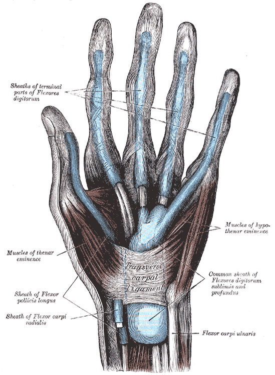
Trigger finger, or stenosing tenosynovitis, is caused by a mismatch between a thickened flexor tendon and the tight fibrous pulley at the base of the finger. The tendon no longer glides smoothly, so the finger catches, clicks or locks in a bent position and sometimes has to be straightened with the other hand. It is more common in women, in people over fifty, and in those with diabetes, an underactive thyroid or inflammatory arthritis.
Typical symptoms
- A tender nodule and pain in the palm at the base of the affected digit
- Clicking, snapping or catching as the finger is bent and straightened, often worst on waking
- Locking of the finger in a bent position, or difficulty bending it at all
- Progressive stiffness if the finger is held in a fixed position for a long time
Management
Recent or mild triggering may settle with activity modification, gentle stretching and a splint that holds the affected joint straight, particularly overnight. An ultrasound-guided corticosteroid injection around the tendon sheath is highly effective and resolves symptoms in a large proportion of patients, with the best results in a single digit of recent onset. Ultrasound confirms the thickened sheath, allows the injection to be placed accurately within it, and avoids injecting into the tendon. A second injection is reasonable if the first gave good but temporary relief.
If triggering recurs after injection, or the finger remains locked, surgical release of the tight pulley gives a reliable and durable cure through a small incision in the palm under local anaesthetic. This short procedure is performed by my hand surgery colleagues.

Ganglion of the wrist and hand
A smooth, jelly-filled swelling arising from a joint or a tendon sheath.
 Click to find out moreClose
Ganglion of the wrist and hand
A smooth, jelly-filled swelling arising from a joint or a tendon sheath.
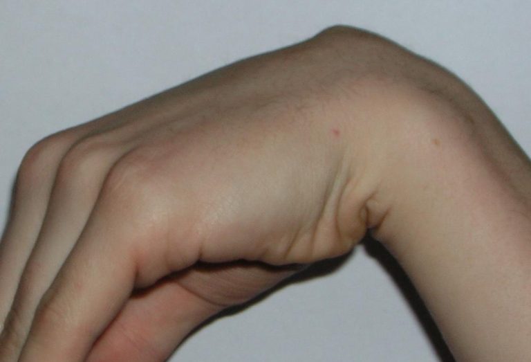
A ganglion is a smooth, firm swelling filled with thick clear jelly-like fluid that arises from a joint or a tendon sheath. The commonest sites are the back of the wrist, the front of the wrist at the base of the thumb, and the base or the last joint of a finger. Ganglia are benign, and many fluctuate in size or disappear on their own.
Typical symptoms
- A visible lump that may become more prominent when the wrist is bent forwards
- A dull ache or discomfort, often worse with weight bearing through the hand or with repetitive activity
- Change in size over weeks or months, sometimes disappearing and returning
- Occasionally pressure symptoms such as tingling if the ganglion lies close to a nerve
How it is managed
Ultrasound confirms the diagnosis, distinguishes a ganglion from other causes of a lump, and shows its origin and its relationship to nearby tendons, arteries and nerves. If the ganglion is painless and the diagnosis is clear, reassurance and observation are entirely reasonable, as a significant proportion resolve without treatment.
Where treatment is wanted, ultrasound-guided aspiration, with or without an injection, can decompress the cyst in clinic and is a straightforward first step, although recurrence is common because the stalk connecting it to the joint remains.
Surgical excision, which I perform myself, is the most definitive option and is advised for ganglia that are painful, recurrent, enlarging, or causing pressure on surrounding structures. The operation removes the cyst together with its stalk and the small cuff of joint capsule from which it arises, which is the key to reducing recurrence. It is normally a day case procedure, and a light dressing and early movement are encouraged afterwards, with most people back to office work within a week or so and to heavier activity after three to four weeks.

De Quervain’s tenosynovitis
A painful, thickened tendon sheath on the thumb side of the wrist.
 Click to find out moreClose
De Quervain’s tenosynovitis
A painful, thickened tendon sheath on the thumb side of the wrist.
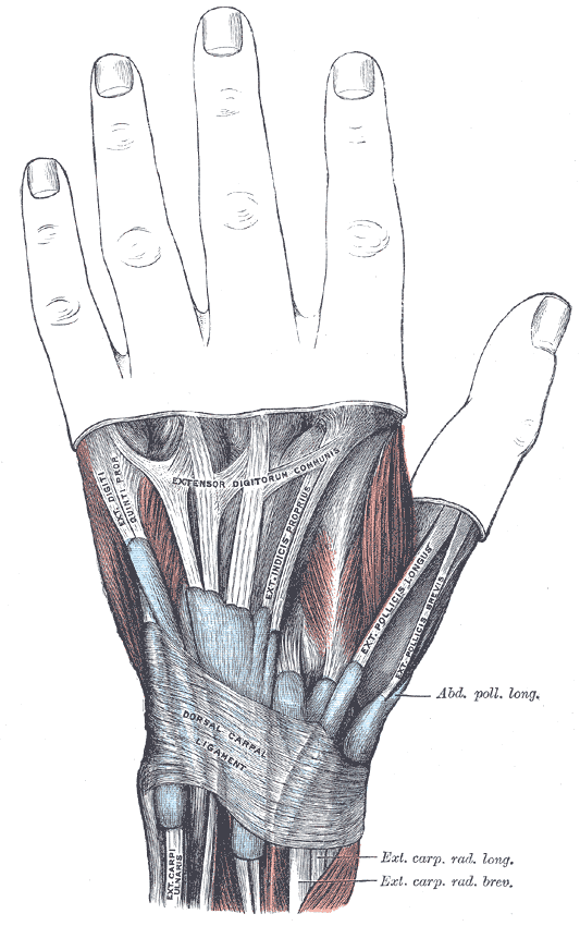
De Quervain’s tenosynovitis is inflammation and thickening of the sheath surrounding the two tendons that run to the thumb on the outer side of the wrist. It causes pain at the thumb side of the wrist that is aggravated by gripping, lifting and wringing movements, and it is particularly common in new parents lifting small children, and after a change in activity or occupation.
Typical symptoms
- Pain and tenderness over the bony prominence on the thumb side of the wrist
- Pain on gripping, pinching, lifting a kettle or turning a door handle
- Swelling and sometimes a creaking sensation over the tendons
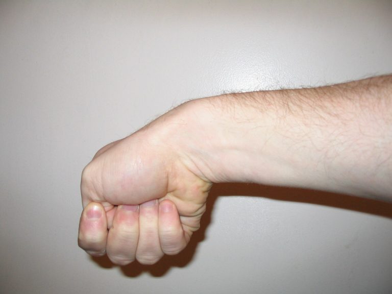
- Pain reproduced when the thumb is tucked into the palm and the wrist bent towards the little finger
How it is managed
First line treatment is a thumb spica splint that rests the wrist and thumb, activity modification, topical or oral anti-inflammatory medication where appropriate, and hand therapy. An ultrasound-guided corticosteroid injection into the tendon sheath is very effective and is the treatment of choice when splinting alone has not settled the pain. Ultrasound is especially useful in this condition because the first compartment is frequently divided by a septum into two separate sub-sheaths; if the injection is placed in only one of them the result is often disappointing, and guidance allows both to be treated.
Surgical release of the first compartment, which I perform myself, is offered when symptoms persist or recur despite splinting and injection. The tight sheath is divided through a small incision on the thumb side of the wrist, taking care to protect the small superficial nerve branches that cross the area, and any septum is released. It is a day case procedure under local anaesthetic. Movement is encouraged straight away, and most people return to light activities within a few days and to full use over three to six weeks.

When to seek advice sooner
- Constant numbness that no longer comes and goes, or visible muscle wasting in the hand
- Rapidly progressing weakness, or a finger or thumb that will no longer straighten
- A hand or wrist lump that is enlarging quickly, hard, or does not transilluminate
- New hand symptoms following significant injury to the wrist or elbow
- Severe pain, spreading redness, fever or discharge from a wound after a procedure
Arranging an appointment
Most hand and wrist conditions can be assessed, explained and treated at a single visit, with diagnostic ultrasound and ultrasound-guided injection carried out at the time of consultation where appropriate. If surgery by a colleague is the better option, that will be discussed openly and arranged without delay. Please get in touch to arrange a consultation.
Contact my PA0333 006 2635
The information on this page is intended as general guidance and does not replace individual medical advice. Please discuss your own symptoms and treatment options with a qualified clinician.